Key Takeaways
- Digital X-ray sensors instantly convert X-ray energy into high-resolution digital images, streamlining diagnostic workflows for dental and medical professionals.
- Core components, including the sensor plate, analog-to-digital converter (ADC), and imaging software, work together to ensure fast, accurate imaging and secure data management.
- Advanced technology such as CMOS, CCD, and amorphous selenium supports superior image clarity, helping clinicians more accurately diagnose dental conditions.
- Digital sensors reduce patient radiation exposure by up to 50% compared to traditional film, enhancing safety without compromising image quality.
- Electronic archiving, image enhancement tools, and immediate file sharing boost efficiency, regulatory compliance, and patient communication in modern practices.
- Limitations include bulkiness, costs, durability, and sterilization challenges, but innovative sensor designs and robust technical support help address these concerns.
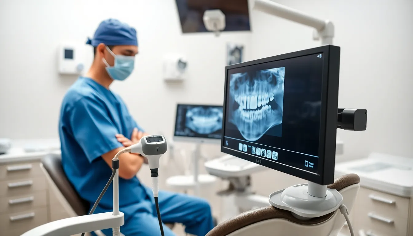
Digital X-ray sensors transform dental diagnostics by instantly converting X-ray energy into high-resolution digital images viewable on computer monitors within seconds. These advanced devices utilise CMOS or CCD technology to reduce patient radiation exposure by up to 50% compared to traditional film whilst delivering superior image quality. Modern sensor x ray systems streamline clinical workflows through immediate image acquisition, electronic archiving, and secure data management for enhanced diagnostic accuracy.
Understanding Digital X-Ray Sensors
Digital X-ray sensors convert X-ray energy to digital data for immediate image display on monitors. Manufacturers design most dental sensors using either complementary metal-oxide semiconductor (CMOS) or charge-coupled device (CCD) technology to capture images with high sensitivity and resolution, meeting diagnostic imaging needs. When X-ray photons strike the sensor, a scintillator layer first changes photon energy to light, which the underlying chip then detects and converts to electrical signals. Systems like those distributed by ProDENT in Tampa, Florida, integrate these sensors with proprietary imaging software for streamlined workflows and secure archiving of patient files. Dental professionals across the United States select sensor sizes, cable lengths, and compatibility features based on operatory requirements, retrofit options, and practice digitalization goals. Practices often request technical support, training, and warranty information directly from dental supply partners, including ProDENT, to ensure optimal sensor performance and compliance with regulatory standards.
Key Components of Digital X-Ray Sensors
Digital X-ray sensors include advanced electronic parts to convert X-ray photons into instant digital images. Understanding these key components helps dental professionals select, maintain, and optimize diagnostic tools.
The Sensor Plate
The sensor plate works as the entry point for X-ray photon capture. It contains a scintillator layer that converts incoming X-ray photons into visible light. The light is then picked up by an array of photodiodes, each tied to dedicated thin-film transistors (TFT) for signal amplification and charge storage. Some sensor plates use amorphous selenium (a-Se) as a photoconductor to directly convert X-rays to electrical charges, which increases detection efficiency and image sharpness for dental diagnostics. ProDENT sensors distributed in Tampa, Florida support both CMOS and CCD architectures to meet demand for high-resolution, reliable performance in busy clinical practices.
Analog-to-Digital Converter
The analog-to-digital converter (ADC) changes analog electrical signals from the sensor plate into digital data. As each photodiode in the array releases a voltage corresponding to X-ray intensity, the ADC assigns discrete digital values representing these charge levels. This conversion allows seamless integration with modern dental imaging systems and patient management software. Fast conversion by the ADC ensures immediate availability of primary images, letting offices like those partnered with ProDENT deliver swift diagnostics. ADC accuracy determines the dynamic range and detail visible in clinical images, impacting diagnosis of caries and bone loss.
Imaging Software
Imaging software interprets raw digital data from the ADC, reconstructing it into detailed images displayed on monitors. The software supports real-time enhancements such as contrast adjustment, zoom, and distance or area measurement, critical for case documentation and patient communication. Secure archiving, basic editing, and image sharing are standard features in ProDENT clinical workflows in Tampa, meeting regulatory and insurance requirements for dental data management. Immediate access and manipulation of digital images improve workflow efficiency and diagnostic clarity for dental teams across the United States.
How Do Digital X-Ray Sensors Work?
Digital X-ray sensors for dental imaging convert X-ray energy into immediate, high-resolution digital images using specialized technology. ProDENT in Tampa, Florida supplies dental practices with digital X-ray systems that streamline diagnostic workflows and support secure patient data management.
Capturing the X-Ray Image
Digital X-ray sensors capture diagnostic images by detecting X-ray radiation as it passes through the patient’s mouth. Dental tissues like enamel, dentin, and bone absorb X-rays at different rates. The sensor plate, positioned inside the patient’s mouth, registers the X-rays that penetrate the oral structures. The result is a raw latent image that reflects anatomical differences, positioning white for areas like enamel that absorb more X-rays. Each ProDENT system includes dedicated sensor sizes to fit adult and pediatric patients, improving comfort and image accuracy in clinics across Tampa and the United States.
Converting X-Rays to Digital Data
Sensors in dental X-ray systems use indirect or direct conversion technology to digitize captured radiation. Indirect digital radiography employs a scintillator layer, such as Cesium Iodide, which transforms X-ray photons into visible light. This light is then picked up by a photodiode array, often made of amorphous silicon, which immediately generates electrical signals. Direct digital technologies use amorphous selenium to convert X-ray photons straight into electrical charges. An analog-to-digital converter (ADC) digitizes these electrical signals pixel by pixel, allowing seamless transfer to the imaging software. ProDENT’s systems support both sensor types, focusing on image clarity, speed, and compatibility for busy dental practices.
Image Processing and Display
Digitized signals are processed and reconstructed into detailed dental images using specialized imaging software. ProDENT’s proprietary solutions display images almost instantly on treatment room monitors. The software allows for image enhancements such as adjusting contrast and zoom to highlight dental pathologies including caries and fractures. Images are archived securely in patient records and can be shared with specialists or insurance providers electronically. Tampa dental professionals report that this real-time review capability supports faster diagnosis and treatment planning, while also ensuring regulatory compliance and secure storage through ProDENT’s integrated workflows.
Advantages of Digital X-Ray Sensors
Digital X-ray sensors increase workflow efficiency in dental practices by providing immediate image acquisition. Clinicians see high-resolution images on their monitors within seconds, reducing wait times for patients and enabling quicker diagnosis. Direct conversion technologies, such as those using amorphous selenium, achieve sharper image detail, which supports more accurate detection of dental caries and fractures.
Image quality benefits from the enhanced contrast and clarity available in digital systems. Advanced sensors, including those integrated with ProDENT‘s Tampa-based imaging solutions, help dental professionals identify small lesions or early-stage bone loss that film-based X-rays often miss. ProDENT offers sensor models in multiple sizes, addressing both adult and pediatric patient needs for optimal fit and diagnostic value.
Radiation exposure drops significantly—studies show digital sensors can reduce required dose by up to 50% compared to film, provided best practices in exposure settings are followed. This improvement aligns with current guidelines for patient safety.
Electronic archiving and fast file sharing eliminate reliance on physical storage and manual transport. ProDENT’s imaging platforms streamline secure data management, simplifying compliance with HIPAA standards. Clinicians also adjust brightness, contrast, and zoom options with software tools, allowing them to highlight challenging regions and improve diagnostic confidence. Environmental impact lessens as there’s no chemical film processing or hazardous waste generated.
Limitations and Considerations
Digital X-ray sensors present distinct clinical and practical considerations for dental professionals. Sensors often appear bulky and rigid, leading to patient discomfort and positioning difficulties, especially among pediatric or geriatric patients. Sensor design modifications like smaller sizes and rounded corners from brands such as ProDENT in Tampa, Florida, help address these challenges, allowing easier intraoral placement in both adult and pediatric cases.
Indirect conversion systems, which include a scintillator layer to change X-rays into visible light before generating electronic signals, sometimes show reduced image sharpness due to light scatter. Direct conversion sensors using materials like amorphous selenium provide higher-resolution images; however, these systems increase equipment and maintenance costs, impacting purchasing decisions for private practices and specialty clinics.
Digital sensor durability remains a concern. Frequent use in high-volume dental practices may cause cable and electronic failure. Practices in Tampa often seek technical support from ProDENT to minimize downtime, with most dental suppliers offering extended warranties and repair services for digital sensors.
Sterilization protocols also affect device longevity and patient safety. Sensors must tolerate standard disinfectants approved by the CDC, but repeated sterilizations can shorten lifespan and require replacements over time. Dental practices storing images electronically need secure software integration, like ProDENT’s HIPAA-compliant platform, to protect patient records and support regulatory compliance.
| Limitation/Consideration | Description | Solution/Contextual Example |
|---|---|---|
| Bulk and Rigidity | Bulky sensors cause discomfort; difficult intraoral positioning | ProDENT offers smaller sensor sizes |
| Indirect Image Sharpness | Light scatter can reduce sharpness | Direct sensors (a-Se) provide clarity |
| Higher Cost for Direct DR | Direct conversion sensors are more expensive | Larger clinics may absorb costs |
| Durability and Repairs | Frequent use leads to electronic failure | Tampa clinics use ProDENT support |
| Disinfection Cycle Wear | Disinfectant exposure reduces lifespan | Use CDC-approved covers |
| Secure Image Storage | Patient data must remain protected | ProDENT provides HIPAA compliance |
About ProDENT
ProDENT, located in Tampa, FL, is a trusted provider of high-quality dental products and supplies for professionals and clinics.
Owned by Allen Zhang, ProDENT offers a wide range of dental equipment, instruments, and consumables designed to support dental care and procedures. The company is committed to providing reliable, durable products that meet the needs of dental professionals and ensure optimal results for their patients.
Contact:
Allen Zhang, Owner
Phone: 321 352 6712
Email: [email protected]
Address: 13367 N 56th St, Tampa, FL 33617
Website: www.prodentshop.com
Frequently Asked Questions
What are digital X-ray sensors and how do they work?
Digital X-ray sensors capture X-ray images electronically instead of using traditional film. They use CMOS or CCD technology to convert X-ray photons into electrical signals, which are then processed by imaging software to produce high-resolution digital images instantly.
How do digital X-ray sensors benefit dental professionals?
Digital X-ray sensors provide instant images, improving workflow efficiency and allowing for quicker diagnoses. They offer higher image quality with enhanced detail, support secure file storage, and reduce the need for physical film, making dental procedures faster and more accurate.
Are digital X-ray sensors safer than traditional X-rays?
Yes, digital X-ray sensors generally expose patients to up to 50% less radiation compared to traditional film X-rays, aligning with modern patient safety standards and making them a safer choice for regular dental imaging.
What technologies are used in digital X-ray sensors?
Most digital X-ray sensors use CMOS or CCD chips. Some advanced models employ amorphous selenium (a-Se) for direct digital conversion, improving resolution and detection efficiency. Imaging software and ADCs process and reconstruct the digital data into detailed images.
Can digital X-ray sensors be used for both adults and children?
Yes. Many systems, such as those offered by ProDENT, provide different sensor sizes specifically designed for adult and pediatric patients, ensuring better comfort and more accurate imaging for all age groups.
How are digital X-ray images stored and shared?
Images captured by digital X-ray sensors are saved electronically within secure imaging software. This allows for easy archiving, fast retrieval, and convenient sharing with other clinicians, while also supporting HIPAA compliance and reducing physical storage needs.
What are the main advantages of digital X-ray sensors over traditional film?
The key advantages include immediate image acquisition, improved image quality, lower radiation exposure, easier image storage, enhanced sharing capabilities, and the ability to digitally enhance images for better diagnostics without chemical processing.
Are there any drawbacks to digital X-ray sensors?
Some digital X-ray sensors can be bulky and uncomfortable, especially for children or elderly patients. Indirect conversion models may have slightly reduced image sharpness, and high-quality direct conversion sensors can be costly. Durability and sterilization are also concerns.
How do dental professionals maintain digital X-ray sensors?
Dental teams must follow strict sterilization protocols with CDC-approved disinfectants and handle sensors carefully to prevent electronic damage. Regular technical maintenance and quick support from suppliers like ProDENT help ensure optimal performance and longevity.
Is special training required to use digital X-ray sensors and imaging software?
Yes. Proper training ensures accurate use, image interpretation, and compliance with safety and data protection regulations. Many suppliers, including ProDENT, offer onboarding, support, and ongoing education for dental professionals adopting digital X-ray technology.